Introduction:
Nuclear medicine is one of the most fascinating and life-changing medical specialties of our time.
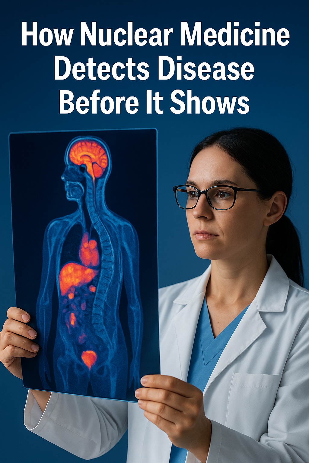
Its unique power lies in seeing what the body hasn’t yet revealed — it visualizes biological processes happening deep inside the body, often before structural changes appear on traditional scans.
By using safe amounts of radioactive substances (radiopharmaceuticals), nuclear medicine can diagnose, monitor, and treat a wide range of conditions — including cancer, neurological, cardiac, inflammatory, and infectious diseases.
What makes nuclear medicine different?
Unlike traditional radiology, which focuses on anatomy, nuclear medicine focuses on function.
This means doctors can see not only how an organ looks, but how it works.
For example, in a kidney study, a small amount of radioactive tracer is injected into the bloodstream. Cameras then track how the kidneys filter the tracer and produce urine. This provides a clear picture of how well the kidneys are functioning, and helps detect blockages or other issues that may not appear on an ultrasound or CT scan.
Key clinical applications
Oncology: Detecting cancer before it’s visible
In recent years, PET/CT and SPECT/CT techniques have revolutionized cancer diagnosis and treatment.
These methods combine functional and anatomical imaging, allowing doctors to detect tumor lesions at early stages and treat certain types of cancer even in advanced stages.
Among the most remarkable advances in treatment:
[^177Lu]Lu-PSMA-617 for advanced prostate cancer (Nature, 2024).
[^177Lu]Lu-DOTA-TATE, effective in treating neuroendocrine tumours (Nature, 2024).
Radium-223, which helps extend survival in patients with bone metastases (Frontiers in Nuclear Medicine, 2024).
Neurology and Cardiology
Neurology, nuclear imaging helps assess brain metabolism, detect early dementia, and locate seizure activity.
Cardiology, it evaluates blood flow and heart function, revealing damaged or underperforming areas.
Inflammatory or infectious diseases, it identifies areas of active biological change even when other imaging results are normal.
Functional imaging vs. anatomical imaging
Type of imaging What it shows Example Anatomical Structure and shape A CT scan shows a visible tumour. Functional (nuclear) Biological activity Detects cancerous cells before they form a visible mass. This combination makes nuclear medicine a powerful tool for early detection, precise diagnosis, and personalized treatment.
Hybrid imaging: PET/CT, SPECT/CT, and PET/MR
Hybrid scanners combine the strengths of both approaches:
Functional imaging (how the body works)
Anatomical imaging (how the body looks)
These integrated systems allow for precise localization of abnormalities, better treatment planning, and improved accuracy.
Newer hybrid machines also offer higher resolution, faster scans, and lower radiation exposure.Theranostics: Diagnosis and treatment in one approach
The newest evolution in nuclear medicine is called theranostics — a blend of therapy and diagnostics.
It uses the same molecular target for both detecting and destroying disease.Here’s how it works:
A diagnostic tracer identifies the target (for example, a tumour).
The same tracer, linked to a therapeutic isotope, is then used to deliver focused radiation to that exact site.
Examples include:
Iodine-131 (I-131) for thyroid disease — used for decades in diagnosis and therapy.
Lutathera (Lu-177-DOTA-TATE) for neuroendocrine tumours.
Pluvicto (Lu-177-PSMA-617) for prostate cancer resistant to hormone therapy (Reuters, 2025).
According to Frontiers in Medicine (2025), there are now more than 30 ongoing Phase 3 clinical trials exploring radiopharmaceutical therapies for various cancers.
Recent breakthroughs in nuclear medicine (2024–2025)
Next-generation radiotracers targeting new molecular markers like PSMA, FAPI, and LAT1 (SpringerLink, 2025).
AI-assisted image reconstruction for higher image quality with lower doses.
Theranostic expansion to include liver, colon, and pancreatic cancers.
Alpha-emitting isotopes such as terbium-161 showing strong results in the Australian VIOLET trial (2025).
Improved accessibility, as more countries develop their own isotopes and nuclear medicine infrastructure.
Challenges and the road ahead
Despite incredible progress, nuclear medicine still faces some challenges:
Limited isotope production and short half-lives.
High equipment and maintenance costs.
Need for trained specialists.
Strict radiation safety and regulatory approvals.
Still, the field’s growth is unstoppable. Nuclear medicine is rapidly becoming a cornerstone of personalized healthcare, combining early detection, targeted therapy, and improved patient outcomes.
Conclusion
Nuclear medicine does more than just diagnose — it saves lives.
By revealing hidden disease processes and delivering therapy directly to affected cells, it bridges the gap between imaging and treatment.As many experts say:
“Nuclear medicine doesn’t just see the body — it sees life inside the body.”
Want to learn more?
👉 Subscribe to our health newsletter and stay updated on the latest medical breakthroughs.
📧 Contact: Contact Form
🔗 Scientific Sources: Nature, Frontiers in Medicine, SpringerLink, Reuters Health